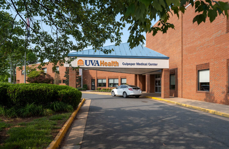
The University of Virginia Health System was first in the nation to image a patient with the next generation of interventional imaging systems, the Artis zeego, on Nov. 14. This news was embargoed until today, the day that Siemens unveils its new technology at the Radiological Society of North America meeting in Chicago. Photos of this first-of-its-kind procedure and video files of the system are available by calling 434-924-9241.
At UVa, this flexible Artis zeego® system helped physicians guide a liver shunt precisely into place using both fluoroscopy and computed tomography (CT slice) imaging. Because of its ease of use, flexibility and speed of imaging, this system has been in daily use for many different patient conditions since that first procedure. UVA has just begun to explore its benefits to patients with different imaging and treatment needs. The system offers versatility, enhanced image quality and streamlined workflow across many clinical environments.
“Because it can quickly and safely image a patient in so many different planes, the zeego system saves us time, which in turn helps to ensure better patient care and safety,” says Dr. Michael D. Dake, chairman of the Department of Radiology at the University of Virginia Health System. The C-arm of this machine is not anchored to the floor or wall, as with other systems, but is mounted onto an articulated robotic arm that allows the C-arm to approach patients from virtually every angle.
“The concept of getting CT-scan-type images in an angiography suite is relatively new, and this system allows us to image the whole abdomen like a conventional CT (computed tomography) and, at the same time, perform complex procedures under fluoroscopic guidance,” said Dr. John F. Angle, an associate professor of radiology and director of interventional radiology at UVA. Fluoroscopy is a form of imaging that uses X-rays, and angiography is the field of radiology that uses X-rays to image blood vessels.
This technology is “ideally suited to chemoembolization of the liver, which is the injection of chemotherapy directly into arteries feeding a liver tumor,” Dr. Angle said.
At UVa, the radiology team now can rapidly compile a 3-D reconstruction from the CT data. Speaking on the day of the first procedure with this system, Dr. Angle said, “What’s exciting is how fast these 3-D images are delivered. These essential reconstructions will be compiled much more quickly.”
The equipment can move in an expanded number of directions to make procedures quicker and minimize the number of times that a patient might need to be repositioned.
The Artis zeego features a multi-axis C-shaped arm that employs robotic technology to extend advanced cross-sectional imaging from head to toe through its virtually unrestricted projection capabilities. Offering a degree of positioning flexibility not achievable with traditional C-arm systems, the Artis zeego allows off-center rotational angiography for all areas of the body.
Again, for photos of the first procedure and of the system, as well as for video of the system, please call 434-924-9241.