CHARLOTTESVILLE, Va. — University of Virginia researchers have developed a revolutionary three-dimensional model that allows them to visualize how breast tissue grows in its earliest stages, giving them the closest look ever at the very beginnings of breast cancer.
This work was described in “Sustained activation of the HER1 – ERK1/2 – RSK signaling pathway controls myoepithelial cell fate in human mammary tissue,” which is published in the August 1, 2011 issue of the prestigious journal Genes & Development . Results from this study and ongoing research at UVA Health System could lead to the development of more effective drugs and even the advancement of personalized medications to treat breast cancer.
The new model represents a major scientific milestone – it’s the first time scientists have been able to successfully and accurately replicate the early growth of human breast tissue outside of the body.
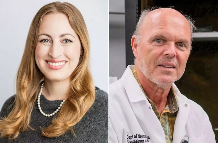
“These findings have important implications for the study and understanding of breast cancer,” says Deborah Lannigan, PhD, associate professor in the UVA School of Medicine ’s Department of Microbiology, Immunology, and Cancer Biology. “Like never before, researchers around the world now have a means of understanding how breast cancer begins and progresses.”
Nearly 90 percent of breast cancers begin in the breast’s ducts, so knowing how they form is crucial to developing improved therapies. A network of breast ducts resembles a tree branch – each arm of the duct is elongated and cylindrical and consists of an outer layer of cells (basal) and an inner layer of cells (luminal). Some breast cancers arise from basal cells and some grow from luminal cells. The outer, basal layer of cells acts as a barrier that is thought to help keep early cancerous cells from escaping out of the ducts and spreading into the rest of the body.
UVA researchers used their model to determine what mechanisms in the body control the development of breast ducts, particularly the outer basal cells. They found that a signal produced by the body, called epidermal growth factor (EGF), tells the ducts how many basal cells to produce. This finding is an important step in understanding how basal tumors might form, and how normal basal cells might prevent the spread of cancers.
Using the new model, UVA researchers also found that human breast tissue is much more sensitive to growth factors than that of mice. “These differences might explain why some drugs are less effective in patients than would have been predicted from animal studies,” suggests Ian Macara, PhD, professor of microbiology and a co-investigator on the study.
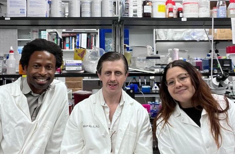
The new model was developed by a unique multidisciplinary team of UVA Health System researchers, including the team of cell biologists Lannigan and Macara, along with breast surgeon David Brenin, MD, and pathologist Chris Moskaluk, MD, PhD. All combined their areas of expertise to devise a novel and better way of studying breast cancer at its earliest stages.
Research funding was supported by the Susan G. Komen Breast Cancer Foundation, the National Institutes of Health as well as local community funds from Patients and Friends of the UVA Cancer Center , and Swing Fore the Cure .