CHARLOTTESVILLE, Va. — Patients with cancer want to know if their chemotherapy is shrinking their tumors. Or if a relative has beginning symptoms of dementia, most families want to know if their loved one has Alzheimer’s disease, so treatment can start as early as possible.
To that end, the University of Virginia Health System has introduced new technology to give UVA patients and their physicians state-of-the-art imaging tools to catch diseases at their earliest stages and design more effective treatments at potentially lower costs.
New cyclotron technology
The centerpiece is a $2 million cyclotron — the first of its kind outside Richmond and the DC area. A circular particle accelerator that is nearly eight feet in diameter and weighs over 50,000 pounds, the cyclotron allows UVA scientists to locally manufacture radioactive elements, which are critical in pinpointing areas of disease.
These radioactive elements are used to turn chemicals, called biomarkers, into imaging agents that accumulate in specific diseased areas of the body. The minute and harmless amounts of radiation allow the biomarkers to appear as bright white spots on a PET (positron emission tomography) scan, telling physicians the exact location of disease.
This imaging technology will benefit UVA patients with such diseases as:
Cancer Alzheimer’s disease Heart disease Drug addiction Traumatic brain injury and other conditions.
Earlier diagnosis, faster treatment
Stuart Berr, PhD, professor of research in radiology and biomedical engineering, and director of the UVA Molecular Imaging Core Lab who spearheaded the cyclotron project, explains the technology’s broad disease applications. Cancer
“One immediate use of this technology will be to assess the effectiveness of a particular cancer therapy,” he says. “You can use chemotherapy and wait months to see if the tumor is smaller. Or we can use PET imaging just days after chemo is administered to see if the treatment is having an effect on the cancer. If not, the therapy can be changed right away. We don’t have to wait for months.”
Heart disease
One advantage of having a cyclotron at UVA is that radiologists will now have access to radioisotopes that decay rapidly, in some cases in as little as two minutes. In those cases, the radioactive elements can be made in the cyclotron in a streamlined process and driven in lead-lined boxes to the UVA Medical Center to be injected into a patient for a PET scan.
“We’ll be able to do ammonia imaging to image cardiac blood flow to see if people have blockages of their coronary arteries,” Berr says. “And we’ll have access to other biomarkers through our partnership with PETNET that we did not have before.” Previously, UVA had to purchase imaging radioisotopes from other facilities.
Alzheimer’s disease
One of the more interesting new imaging biomarkers is called Florbetapir, marketed by Eli Lilly and Company, that can be used to image Alzheimer’s disease since it binds to the plaques found in patients with the disease. The problem is that the imaging biomarker decays rapidly. “So it will be very useful be able to make these short-lived biomarkers and get them to the medical center quickly,” Berr says.
The technology, located at UVA’s Sheridan G. Snyder Translational Research Building at Fontaine Research Park, was purchased through a 2008 grant from the National Institutes of Health and is operated in partnership with PETNET, a subsidiary of Siemens Medical Solutions. Dr. Berr served as principal investigator of the grant. The entire project took more than five years to complete at a total cost of some $4 million.
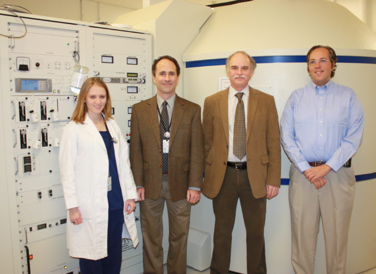
Pictured (left to right) with the cyclotron are:
Darci Sidwell, RPh, PharmD, BCNP, PETNET facility manager; Stuart S. Berr, PhD, professor of research, Radiology and Biomedical Engineering, director of the UVA Molecular Imaging Core Lab, and principal investigator of the NIH grant used to purchase the cyclotron; Brent A. French, PhD, associate professor of biomedical engineering, cardiovascular medicine, and radiology; and James R. Stone, MD, PhD, assistant professor of radiology and medical imaging.
Other faculty involved with grant proposal (not pictured):
Frederick H. Epstein, PhD, professor and chairman, Department of Biomedical Engineering; Eric Houpt, PhD, associate professor of medicine, Division of Infectious Diseases; Bankole A. Johnson, DSc, MD, PhD, MPhil, FRCPsych, chairman, Department of Psychiatry and Behavioral Sciences; Victor Laubach, PhD, associate professor of research, Department of Surgery.