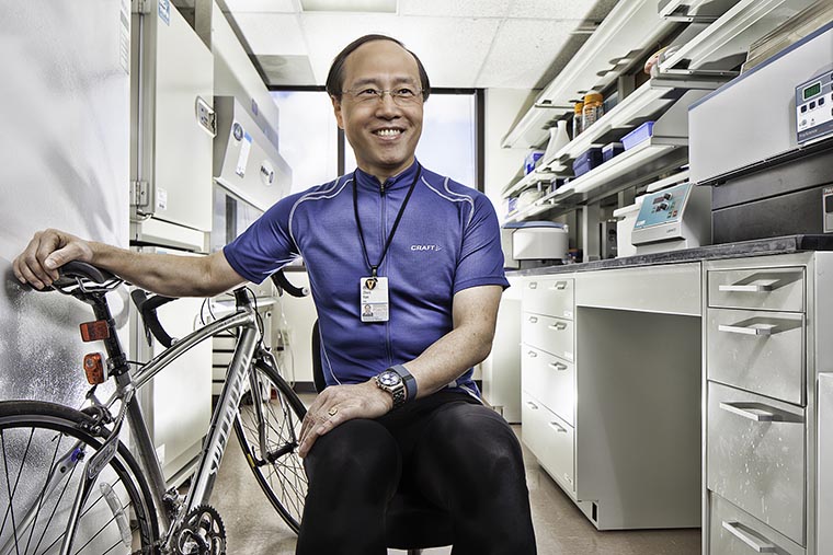
Zhen Yan, PhD, is a top exercise researcher. Pictured with his bike, he practices what he preaches.
A top exercise researcher at the School of Medicine has revealed how our bodies ensure the proper functioning of the powerhouses of our cells. The findings could open the door to better treatments for many common diseases, including Alzheimer’s and diabetes.
The new research from UVA’s Zhen Yan, PhD, and colleagues reveals how our cells sense problems and perform quality control on cellular “batteries” known as mitochondria. Yan has spent many years seeking to better understand the workings of mitochondria, and he calls the new discovery the most exciting of his career.
“Mitochondria are the center of universe to me since literally all cells in our body rely on mitochondria for energy production and must have a bulletproof system to ensure the powerhouses are functioning properly,” said Yan, the director of the Center for Skeletal Muscle Research at UVA’s Robert M. Berne Cardiovascular Research Center. “Chronic diseases, also known as noncommunicable diseases, such as diabetes, heart failure and Alzheimer’s disease that catastrophically impact so many individuals, families and the whole society are caused by problems of the mitochondria in the cells.”
Stress Detectors
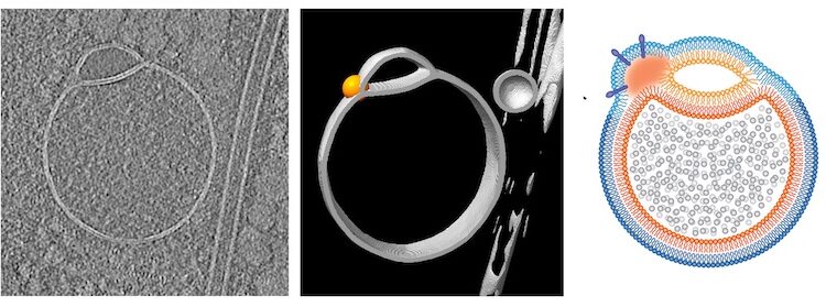
Yan and his team discovered special sensors on the outer membrane surrounding the mitochondria in various tissues in both mice and humans. These sensors detect “energetic stress,” such as caused by exercise or fasting, and signal for damaged mitochondria to be degraded and removed. This essential cleanup process is known as “mitophagy,” and its existence was first suggested more than 100 years ago. But how it works has never been fully understood. Yan’s new research offers long-sought answers.
Yan and his colleagues found that the mitochondrial sensors, known as “mitoAMPK,” exist in slightly different forms in different tissues. For example, one type seemed particularly active in skeletal muscle. In a new scientific paper outlining their findings, the researchers describe the variety of sensors as “unexpectedly complex.” They go on to outline how these sensors provide a vital damage-control system that safeguards our cellular energy supply.
One finding of the study that Yan finds extremely exciting: Treating mice with metformin, the most effective, first-line anti-diabetes drug, activates mitoAMPK in skeletal muscles without activating AMPK in the other parts of the cells. The finding is the best illustration of the importance of activating mitoAMPK and mitochondrial quality control in treatment of a common chronic disease that is known to be caused by accumulation of dysfunctional mitochondria in our body. It also explains why regular exercise is so powerful in preventing and treating such diseases.
The new insights gained into mitochondrial quality control will boost efforts to develop new treatments for non-communicable diseases that have reached pandemic proportions and are estimated to cause 71% of all deaths.
Yan, who is part of UVA’s Division of Cardiovascular Medicine, says it will be important for doctors to better understand how specific diseases interfere with mitochondrial function. And his new findings set the stage for that.
“We have developed genetic models for pinpointing the key steps of mitoAMPK activation and are on our way to discover the magic molecules that are controlled by mitoAMPK,” Yan said. “The findings taught us a lot about the beauty of the sensor system in our body. Society should definitely take advantage of these findings to promote regular exercise for health and disease prevention and develop effective exercise-mimetic drugs.”
Mitochondria Findings Published
The researchers have published their findings in the scientific journal PNAS. The research team consisted of Joshua C. Drake, Rebecca J. Wilson, Rhianna C. Laker, Yuntian Guan, Hannah R. Spaulding, Anna S. Nichenko, Wenqing Shen, Huayu Shang, Maya V. Dorn, Kian Huang, Mei Zhang, Aloka B. Bandara, Matthew H. Brisendine, Jennifer A. Kashatus, Poonam R. Sharma, Alexander Young, Jitendra Gautam, Ruofan Cao, Horst Wallrabe, Paul A. Chang, Michael Wong, Eric M. Desjardins, Simon A. Hawley, George J. Christ, David F. Kashatus, Clint L. Miller, Matthew J. Wolf, Ammasi Periasamy, Gregory R. Steinber, D. Grahame Hardie and Yan.
The research was supported by National Institutes of Health grants R01-AR050429, R00-AG057825, R01-AG067048 and T32 HL007284-37; American Heart Association post-doctoral fellowship 14POST20450061 and grant 114PRE20380254; Canadian Institutes of Health Research Foundation Grant 201709FDN-CEBA-116200; Diabetes Canada Investigator Award DI-5-17-5302-GS; and a Tier 1 Canada Research Chair and the J. Bruce Duncan Endowed Chair in Metabolic Diseases. As part of their work, the researchers used a UVA Keck Center Zeiss 780 multiphoton FLIM-FRET microscope and Leica SP5X confocal supported by the NIH.
To keep up with the latest medical research news from UVA, subscribe to the Making of Medicine blog.