School of Medicine researchers have received $3.1 million from the National Institutes of Health’s National Cancer Institute to pioneer the use of focused soundwaves to improve treatment of debilitating cerebral cavernous malformations (CCMs).
These vascular lesions, often called cavernomas, are overgrowths of tiny blood vessels called capillaries in the brain or spinal cord. The growths can cause seizures, severe headaches, paralysis and more. If they trigger bleeding in the brain, they can be deadly.
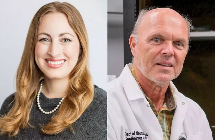
Surgery, when possible, remains the primary treatment option for symptomatic cavernomas. But UVA Health researchers led by Richard J. Price, PhD, and Petr Tvrdik, PhD, are seeking to create a powerful new method to deliver drugs to the lesions. Their approach combines the power of focused ultrasound with tiny microbubbles to allow drugs to reach lesions and the surrounding “microenvironment” in a way now impossible.
“Many CCM patients are in desperate need of more-effective treatment options. Some patients have surgically inaccessible lesions that can only be treated with radiation, but such treatments can have strong side effects and take a long time to show efficacy,” said Price, co-director of UVA Health’s Focused Ultrasound Cancer Immunotherapy Center. “We ultimately wish to treat these lesions non-invasively with biologic drugs and gene therapies, but these therapies are relatively large in size and do not penetrate well into brain tissue. Low-intensity focused ultrasound can be steered almost anywhere in the brain. It gives us a unique opportunity to precisely deliver such advanced therapies right to the CCM.”
About Cerebral Cavernous Malformations
One in 500 people are thought to have at least one cavernous malformation. In a majority of cases, the lesions cause no problems, but for many patients, they trigger symptoms that worsen as the blood vessels continue to grow. For some people, these symptoms can become debilitating or even fatal.
One of the great challenges in treating cavernomas lies in their inaccessibility for both surgical intervention and medication. Brain surgery can be risky; drugs, on the other hand, often can’t reach the malformations because of the brain’s natural protective barrier, known as the blood-brain barrier. This barrier is designed to keep harmful substances out, but it also serves as a blockade to many therapeutic treatments. This is especially true for larger “biologic” drugs derived from living organisms.
Price, of UVA’s Department of Biomedical Engineering, and Tvrdik, of the Department of Neurosurgery, are aiming to briefly breach that protective barrier in a very precise way. Their approach uses microbubbles in conjunction with focused sound waves to open, temporarily, the blood-brain barrier exactly where it is needed. This can allow drugs to penetrate where they normally cannot. Once the drugs are in, the barrier is allowed to close and resume its normal function.
“Our clinically relevant mouse model demonstrates that cavernomas contain stagnant blood pools, mirroring the human condition,” Tvrdik said. “Interestingly, the effects of focused ultrasound alone proved to be quite beneficial, with no adverse complications.”
Price and Tvrdik have already had promising success testing their approach in their genetically faithful mouse model of the disease. Remarkably, they found that the sound wave-microbubble combo alone was enough to stabilize CCMs even without medication. That suggests that there could be additional benefits to the approach above and beyond allowing drug delivery.
The researchers will use their five-year NCI grant to develop an image-guided system that will let doctors watch as the blood-brain barrier is opened, so that they can ensure medicines are delivered exactly where needed. The researchers will then seek to better understand how these drugs can be optimized to control, and perhaps even shrink, the cavernomas.
Ultimately, they hope to be able to control the growth of the lesions without the need to cut into the skull. If successful, that could transform how cerebral cavernous malformations are managed and treated.
“Given our outstanding focused ultrasound infrastructure and neurosurgical expertise at UVA, I am hopeful that we can translate successful pre-clinical findings into trials fairly soon,” Price said. “I continue to be fascinated by how we can safely, and non-invasively, manipulate tissue and blood vessels in a controlled manner with sound waves. Fingers crossed this is yet another disease indication that will benefit from focused ultrasound technology.”
About Focused Ultrasound
UVA Health was one of the earliest pioneers in the field of focused ultrasound. Research by UVA’s Jeff Elias, MD, and colleagues has already paved the way for the federal Food and Drug Administration’s approval of the technology to treat Parkinson’s disease symptoms and essential tremor, a common movement disorder. UVA’s expertise with the technology has led to a robust research program that is examining the use of focused ultrasound to treat many conditions, including cancer.
The tremendous promise of focused ultrasound prompted UVA Health and the Charlottesville-based Focused Ultrasound Foundation to launch the Focused Ultrasound Cancer Immunotherapy Center, the world’s first center dedicated specifically to advancing a focused ultrasound and cancer immunotherapy treatment approach that could revolutionize cancer care for the 21st century.
Learn more about about focused ultrasound at UVA.
The CCM research is funded by NCI grant No. R01CA279134.
To keep up with the latest medical research news from UVA, subscribe to the Making of Medicine blog.